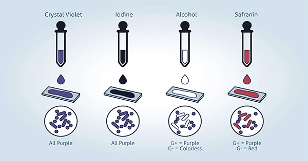
Based on the color of the stain, the Gram stain is a differential staining technique that identifies which bacteria are Gram-positive or Gram-negative. One reagent used in this technique to achieve color distinction is acetone alcohol. Gram-negative germs have little to no peptidoglycan layer and stain pink, while gram-positive bacteria have a thick peptidoglycan layer and stain purple.
Primary Stain-Crystal Violet: After preparing a slide with a bacterial sample, crystal violet is used to stain the piece. Both Gram-positive and -harmful bacteria will now have a purple appearance. The slide is typically stained with crystal violet for 30 seconds before any extra stain is removed with water. Because the crystal violet can slightly bind to the peptidoglycan layers, some of the primary stains will remain after washing with water.
Mordant-Iodine: The sample is then exposed to iodine for a minute. It functions as a mordant, fixing dyes during the staining process. When iodine binds to crystal violet, an insoluble complex is formed that sticks to the thick peptidoglycan coating seen on Gram-positive bacterial cells considerably better than crystal violet alone does. After applying the iodine, there is no water-washing step.
Decolorizer-Alcohol: The decolorizing agent might be either acetone or ethyl alcohol. The crystal violet-iodine combination can escape through the weaker peptidoglycan layer when the alcohol dissolves lipids in the outer cell membrane of Gram-negative bacteria. Once all of the iodine has been rinsed away and the run-off is colorless, the alcohol is added for 10 to 20 seconds before being poured on the slide. At this stage of the Gram stain procedure, Gram-positive bacteria still contain crystal violet while Gram-negative bacteria are colorless. To stop the decolorizing effect, the slide must be cleaned with water after use.
Counterstain-Safranin: Then, safranin is included to improve contrast and visibility with the colorless Gram-negative bacteria. Under a microscope, the stain gives these bacteria a pink appearance. Gram-positive bacteria are also stained by the stain because it is added to the entire sample, but the deeper crystal violet masks the paler safranin pink color. After the slide sample has been saturated with safranin for roughly a minute, any excess stain that did not stick to the bacterial cells is washed off with water.